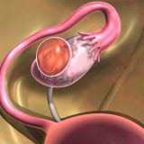
A colloidal cyst is a benign tumor that grows slowly in the human brain. The cystic mass is filled with colloidal (gelatin) fluid encapsulated by connective tissue cells. A colloid cyst can appear not only in the brain, but also in the patient’s thyroid gland.
Colloid cyst in the thyroid gland and brain – the difference in the development of the disease
- During the formation of a colloid cyst in the brain, the circulation of fluid flows is blocked. Because of this violation, the development of hydrocephalus starts. Also, constant pressure of the cystic formation on other parts of the brain can lead to sudden death;
- A colloidal cyst in the thyroid gland has a more favorable course. Most often, these benign tumors contribute to the development of an abscess and inflammation of certain areas.
The main treatment for colloid cysts is surgery.
What causes a colloidal cyst?
Medical experts still could not give an exact answer about which cells in our body cause the development of colloidal cysts of the brain. However, they found that cystic formation occurs at the stage of embryogenesis during the formation of the central nervous system of the child. Such a cystic formation may not show symptoms until the person is fully grown. Usually by this age, the cyst reaches such a large size that it begins to put pressure on the patient’s organs and tissues.
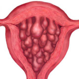
The formation of a colloid cyst is most often associated with hyperplasia and the degeneration of its follicles.
The main causes of the appearance of a colloid cyst:
- Lack of iodine in the human body;
- Sleep disturbance;
- Uneven daily routines;
- Tobacco smoking;
- Constant neuro-emotional stress;
- Frequent pregnancy;
- Permanent stay in a cold space (living in the northern regions);
- X-ray irradiation.
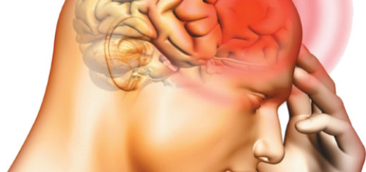
Symptoms of a colloid cyst
Most often, a colloidal cyst does not cause discomfort in the patient. However, when a cystic formation begins to increase sharply, a person experiences a sharp deterioration in health status:
- Persistent headaches;
- Vomiting;
- Loss of consciousness;
- A sharp decrease in visual acuity;
- Ringing in the ears;
- Mental personality disorders;
- Incontinence;
- Loss of coordination;
- Tremor.

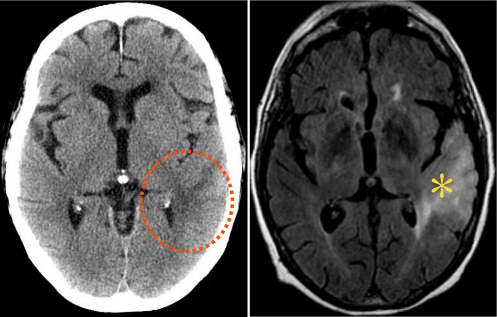
Diagnosis of colloid cyst
When diagnosing a colloid cyst, the method of magnetic resonance imaging is used. In rare cases, it happens that the cyst is an isodenzated MRI of the brain. In this case, the most effective diagnostic method is computed tomography. Thanks to MRI of the brain, one can easily determine the location of the colloid cyst and its location relative to the cerebral structures of the brain.
During brain MRI, a benign tumor is detected. Usually it is located in front of the third ventricle. At this point, the cystic formation blocks the opening of the monroe, which triggers the expansion process of one of the lateral ventricles.
How is a colloidal cyst of the brain treated?
Unlike a colloid cyst of the thyroid gland, a colloidal cyst of the brain cannot be left without attention and surgical intervention. If you use traditional methods and means as a treatment, then this will not affect the state of cystic formation.
The presence of a colloidal cyst of the brain is accompanied by constant pain and a deterioration in the general condition of the human body. Unfortunately, to date, medical experts have not yet been able to find an effective method of surgery that would help get rid of a cyst without consequences.
Surgical Options
Despite the fact that there is no most effective method of surgery for the treatment of cystic education, doctors have found 4 safe ways to remove a benign tumor:
- Transcallose access – performed by craniotomy of the occipital region of the head. Most often, the right hemisphere of the patient is selected, in which the left hemisphere of the brain is dominant;
- Transcortical access – carried out through the middle temporal gyrus;
- Stereotactic access – a neurosurgical method of brain surgery;
- Ventriculoscopic removal – an endoscopic method for examining the brain.
How is the operation performed?
Usually, an operation to remove a cystic mass is performed using the endoscopy method. Surgeons gain access to the hemisphere and hemispheres of the patient’s brain.
If the cyst grows repeatedly, the surgeon prescribes a second operation, where a puncture and emptying of the cavity of the cystic formation are performed. Later, a sclerosant is introduced into the cavity of the colloid cyst, because of which its walls are glued together.
A colloid cyst can be a harmless benign tumor or a dangerous disease that can lead to death. It depends on the location of the cystic mass. But you should remember only one thing: if the cyst begins to grow sharply and causes you great discomfort, then the only treatment is surgical intervention!