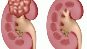
A complex kidney cyst is characterized by the presence of partitions between the cystic cavities. It is a multi-chamber cyst. It must be carefully examined, because it often develops into a malignant formation. In most cases, the treatment of complex kidney cysts requires surgery.
Causes of complex kidney cysts
A complex kidney cyst can develop as a secondary manifestation of a simple cyst. Such relapse occurs as a result of complications such as infection or bleeding, and also as a result of the proliferative process (tissue growth due to pathologically active cell division).
As is the case with simple kidney cyst formation, different types of complex kidney cyst can give similar pictures on medical ultrasound diagnosis. Therefore, it is not possible to make an accurate diagnosis without additional examinations.
However, the timely detection of a complex kidney cyst with using ultrasound is already important in the successful treatment. Features indicating the presence of cystic formation during ultrasound diagnostics:
– thickening and irregular contour of the cyst wall (segmental or along the entire length);
– presence of partitions;
– presence of calcification;
– the presence of seals or solid elements in the tissues present in the plural;
– distinct vascularization (formation of new blood vessels).
Complex kidney cyst complications
Complex kidney cyst has its complications. It’s important not to allow starting of inflammatory processes. They often occur in the fluid filling the cyst cavity.
In addition, there may be a hemorrhagic kidney cyst. This occurs when there is bleeding inside the cyst. Blood partially or completely fills the cavities of a complex kidney cyst.
Echinococcosis cyst is extremely unpleasant thing caused by hydatid tape worm. This disease is called hydatidosis.
As a rule, mature worm lives in the small intestine of a dog. Man is a random intermediate “master”. The most common localization site is the liver. Renal hydatidosis is about 2% of human echinococcal cysts.
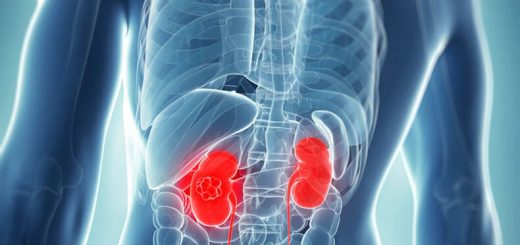
Renal cancers account for about 7-10% of cystic lesions detected by ultrasound. Although the percentage is small, the risk of cancer is worth carrying out some additional diagnostic procedures to make accurate diagnose.

