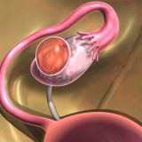
Liver cysts are in most cases harmless changes that do not cause serious symptoms and are most often detected by chance with diagnostic imaging. Liver cysts are rarely associated with the development of liver cancer. Large and numerous cysts on the liver may exhibit different symptoms:
- feeling of pressure and satiety in the stomach;
- discomfort in the abdomen;
- bloating.
What are liver cysts?
Liver cysts are quite rare and are usually detected by chance during the visualization of the abdominal cavity. Most liver cysts do not cause discomfort, which would cause the patient to see a doctor. Symptoms occur when the cyst reaches a significant size or when it is pressed against other structures due to its location.
A cyst in the liver is usually a mild disease that does not pose a threat to the patient’s health. It may, however, be caused by a parasitic infection or cancer. Therefore, it requires specialized treatment.
Causes of liver cysts
The most common type of cysts located in the liver are congenital changes. In adult patients, this is usually a single outbreak, while polycystic liver disease is in most cases associated with the simultaneous appearance of multiple cysts in the kidneys. Causes of liver cysts:
- congenital changes, most often resulting from disorders in the development of bile guides in the life of the fetus;
- genetic determinants that cause the development of polycystic diseases of the liver and kidneys;
- abdominal trauma, contributing to the development of post-traumatic cysts;
- liver cancer, especially primary liver metastases;
- infection by parasites, especially tapeworms;
- Diseases of the intrahepatic bile ducts.
The process of diagnosis and treatment of a patient with a cyst on the liver depends largely on the cause of the cyst, so it is important to determine it at an early stage of treatment.
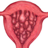
Types of liver cysts
Congenital cysts
A congenital liver cyst is a change that can vary in size. There are cystic formations from small diameters to large sizes, but usually they do not exceed 10 cm. They are surrounded by a thin-walled bag, and the inside is filled with a brownish liquid. A characteristic feature of cystic liver disease is a significant number of changes that increase with the patient’s age.
Physical injury
As a result of blunt abdominal damage, post-traumatic cysts are formed, which most often contain blood, bile or necrotic tissue. Because of their design, they are called pseudo-cyst cysts.
Liver cancer
Very rarely, the cause of liver cysts is cancer. Cystadenoma (benign tumor) or cystadenocarcinoma (malignant tumor) can develop inside the cyst. Both diseases are most commonly associated with middle-aged women. As a rule, they are filled with mucous fluid.
Infestation by parasites
Large cysts containing several liters of fluid develop as a result of infection with tapeworm, most commonly Echinococcus granulosus. Characteristic of the changes caused by this parasite are calcification of bags with cysts, which are often seen during X-ray examination.
Symptoms of cyst
Liver cysts show symptoms only when they reach a considerable size.
- Large or numerous changes usually cause a feeling of pressure or fullness in the epigastric or lower chest;
- The patient experiences discomfort that sometimes cannot be detected;
- Bloating and vomiting may occur if the stomach is constricted;
- Cancer changes are usually accompanied by general symptoms in the form of lack of appetite, weight loss or bad mood;
- There is an itch that occurs during infection about a cystic cyst;
There is a risk of a cyst rupture, the contents of which may leak into the abdominal cavity. This is due to the appearance of sudden, acute pain. Anaphylactic shock or peritonitis may also develop.
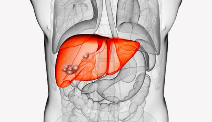
Diagnosis of liver cysts
Liver imaging plays a crucial role in the diagnosis of liver cysts. Liver cysts are visualized with ultrasound, computed tomography or magnetic resonance imaging. They are also useful in assessing the location, size, and structure of changes, which then influence the choice of treatment.
A typical liver cyst manifests itself in ultrasound in the form of a round smooth-wall fluid reservoir. A thick wall of lesions, the presence of calcifications, septa in the lumen of the cyst may indicate the occurrence of hydatid cyst. In the case of congenital cysts, biochemical blood tests are usually normal, unless, of course, the patient has another state at the same time.

Treatment of cysts
Therapy for liver cysts depends on the cause of the change. Large congenital cysts that cause symptoms are indications for surgery. Most often, part of the wall of the lesion is dissected (so-called fenestration), which is then subjected to histopathological examination. The fluid filling the cyst undergoes bacteriological examination.
In case of polycystic liver, fenestration of the largest cysts is carried out to reduce the symptoms, in some cases it is necessary to isolate part of the liver parenchyma. Liver transplantation is very rarely necessary. Tumor cysts are removed along with part of the liver parenchyma during surgery. Detection of a cryptopharyngeal cyst that does not give symptoms is an indication for quick treatment, because there is a risk that the change can become more rapid. As a rule, a cyst is enucleated or a part of the liver is removed.